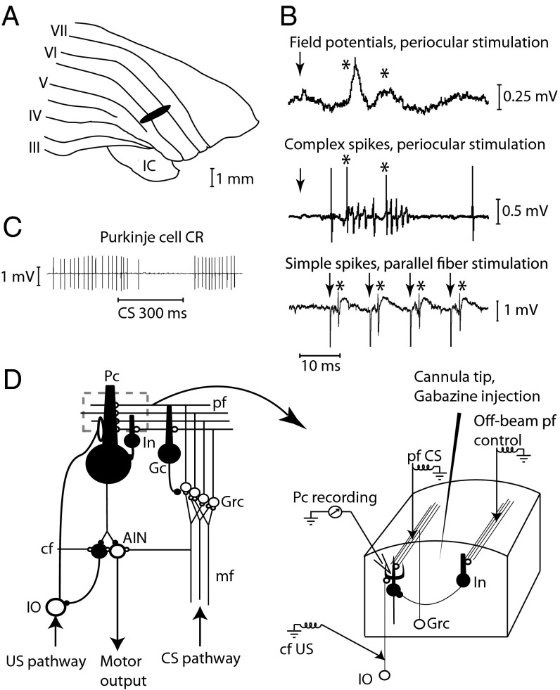
Fig. 1.
Experimental setup. (A) Blink-controlling area in cerebellar cortex. IC, inferior colliculus; Roman numerals indicate cerebellar lobules. (B) Periocular stimulation (1 pulse, 300 μA) elicits short-latency field potential responses on the cerebellar surface. Below are single-cell recordings of two complex spikes elicited by the periocular stimulation (1 mA) and simple spikes elicited by parallel fiber stimulation (4 μA). Arrows indicate stimulation; asterisks indicate responses. (C) Typical conditioned Purkinje cell response (CR). (D) Neuronal wiring diagram with stimulation, recording, and injection sites. AIN, anterior interpositus nucleus; CS, conditional stimulus; cf, climbing fiber; Gc; Golgi cell; Grc, granule cells; In, interneuron; IO, inferior olive; mf, mossy fibers; Pc, Purkinje cell; pf, parallel fibers; US, unconditional stimulus.

