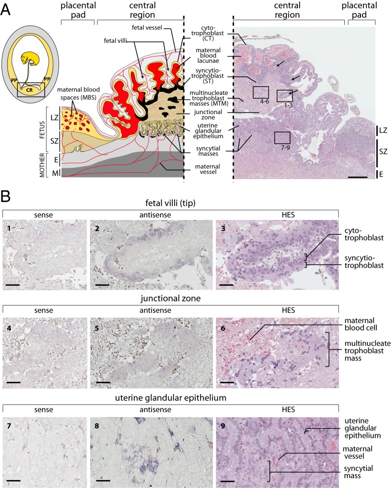
Fig. 6.
Structure of the S. setosus placenta and in situ hybridization for syncytin-Ten1 expression on placental sections. (A) Schematic representation and hematoxylin eosin saffron (HES)-stained section of a midgestation S. setosus placenta. (Left, Inset) Overview of a gravid uterus displaying the hemophagous central region (CR) and the peripheral placental pad (PP) of the placenta. The yellow and gray areas represent the fetal and maternal tissues, respectively. (Left) Detailed scheme of the midgestation placental structure, with the maternal and fetal vessels schematized in red. In the placental pad tissues are, from mother to fetus, the myometrium (M), the endometrium (E), the spongy zone (SZ), and the labyrinthine zone (LZ) with cytotrophoblasts (yellow) delineating maternal blood spaces (MBS). The structure of the central region is highly divergent from that of the placental pad, with hemophagous columnar CTs (yellow) forming villi delineating large maternal blood lacunae. A thick ST layer (black) can be observed at the tip of the villi contacting a junctional zone. This junctional zone is composed of a meshwork of degenerating tissues through which maternal blood diffuses to the lacunae and in which large multinucleate trophoblast masses (MTM) are present (also represented in black). The maternal glandular epithelium in the central region is well preserved, and the ST contacts it only at the periphery. Syncytial masses also are present underneath the glandular epithelium. (Right) HES-stained section of a midgestation placenta with the positions of panels 1–9 in B indicated. Fetal villi are indicated by arrows. (Scale bar: 500 µm.) (B) In situ hybridization using digoxigenin-labeled antisense or sense riboprobes on serial sections of the HES section shown in A from the central region of the placenta revealed with an alkaline phosphatase-conjugated anti-digoxigenin antibody. (1–3) Tip of a fetal villus covered with a ST layer contacting the junctional zone. The ST is specifically labeled with the antisense probe. (4–6) Detail of the junctional zone centered on a multinucleate trophoblast mass that appears faintly labeled by the antisense probe. (7–9) Detail of the uterine glandular epithelium with the intrauterine syncytial masses, also specifically labeled. (Scale bars: 50 µm.)

