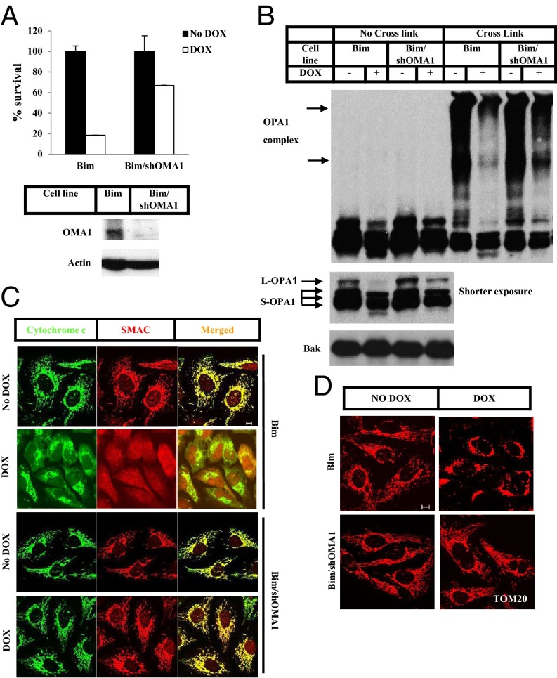
Fig. 2.
OMA1 controls Bim-induced OPA1 cleavage and disassembly of the OPA1-containing complex. (A) U2OS_Bim (Bim) cells and U2OS_Bim cells stably expressing OMA1 shRNA (Bim/shOMA1) were treated with or without Dox for 12 h. Cell viability was determined using the Cell-Titer Glo kit. Whole-cell extracts of Bim and Bim/shOMA1 cells were analyzed by Western blotting. (B) Bim and Bim/shOMA1 cells were treated with or without Dox for 8 h. The P15 fractions of the cells were cross-linked with 10 mM BMH where indicated. Western blotting was performed using the indicated antibodies. (C and D) Bim and Bim/shOMA1 cells were treated with or without Dox for 8 h. z-VAD was included during the treatment of Bim cells. Immunostaining was performed using anti-cytochrome c (green) and anti-Smac (red) antibodies (C) or anti-TOM20 antibody (D). (Scale bars: 10 µm.)

