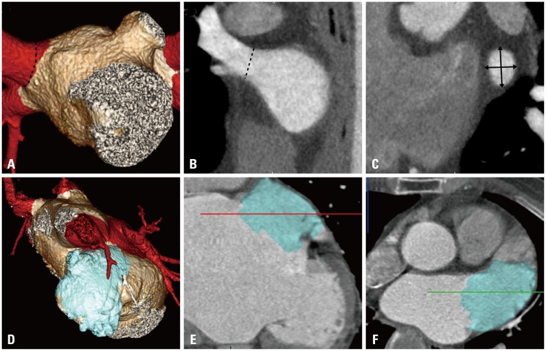
Fig. 1.
Multiplanar reconstructed (MPR) images illustrating the double-oblique measurements of pulmonary veins (PVs) and left atrial appendage (LAA) ostial diameters. (A) 3D reconstruction image of left atrium and pulmonary veins. The dotted line denotes right superior PV ostium. (B) Oblique MPR view of the right superior PV. The ostium (dotted line) was confirmed in multiple views. (C) Enlarged axial MPR view across the ostium of the superior PV showing measurements of the PV diameter (black arrows). (D) 3D reconstruction image of LAA. (E and F) MPR view of the LAA showing enlarged LAA. 3D, three-dimensional.

