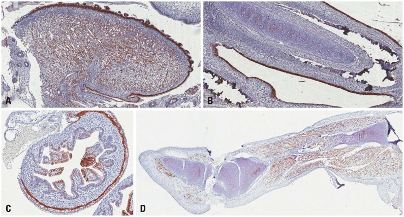
Fig. 3.
The immunohistochemical analysis of the enteroviral VP1 capsid protein from tissues in an encephalocele case. VP1 was highly expressed in the epithelium and muscle layer of the tongue (A) and respiratory epithelium (B), mucosal and muscle layer of the small intestine (C), and leg muscles (D).

