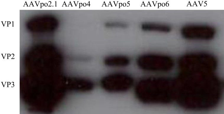
Figure 2. Porcine-derived AAVs form VP1, VP2, and VP3.
DNA encoding the cap of porcine-derived AAVs was transfected into HEK293T cells. Cell lysate was harvested and western blot performed using monoclonal antibody against AAV VP1, VP2, and VP3 (Fitzgerald). Western blot reveals that porcine-derived AAVs form VP1, VP2, and VP3 with sizes of ~87, 72, and 62 kiloDaltons, respectively. AAV5 VP1, VP2, and VP3 were used as a comparison.

