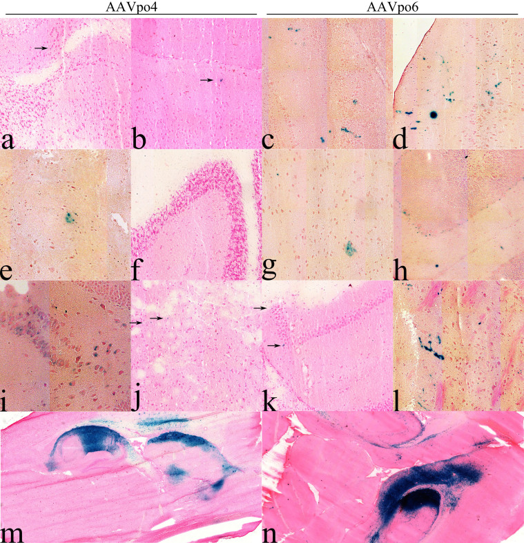
Figure 8. Porcine-derived AAVs transduce cells in the mouse brain.
AAVpo4-LacZ and –po6-LacZ were assessed for their ability to transduce cells in the CNS of BALB/c mice following IVTV or IC administration of AAVpo4-LacZ or –po6-LacZ at a titer of 1 × 10¹¹ gc/mouse. Following IVTV injections, AAV-LacZ transduction was observed in: (a, c) olfactory bulb, (e,g) striatum, (i, k) hippocampus, (b,d) cortex, (f, h) cerebellum, and (j, l) medulla of the mouse brain. (m, n) Cross sections of mouse brains injected IC with AAV-LacZ vectors. Mouse brains were frozen with O.C.T. and sectioned using a cryostat at 10 μm. Each section was stained with X-gal and eosin or nuclear fast red stain. Blue cells represent AAV-LacZ transduced cells (anecdotal transduction is indicated by black arrows).

