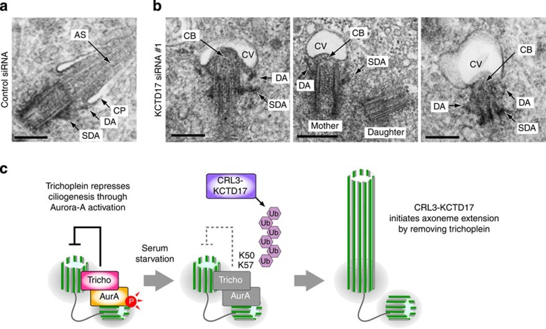
Figure 5. KCTD17 depletion blocks axonemal extension during ciliogenesis.
(a,b) Transmission electron micrographs of mother centrioles (single sections) in PRE1 cells transfected with control (a) or KCTD17 siRNA #1 (b), followed by 24 h serum starvation for ciliogenesis. AS, axonemal shaft; CB, ciliary bud; CP, ciliary pocket; CV, ciliary vesicle; DA, distal appendages; SDA, subdistal appendages. Scale bars indicate 500 nm. (c) Proposed model.

