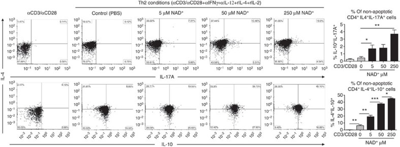
Figure 6. NAD+ promotes IL-10 and IL-17A in Th2 polarizing conditions.
Sorted naïve CD4+ T cells were isolated from spleens of 5C.C7 Rag2−/− mice (C57BL/6 background) and were activated with α-CD3/α-CD28 antibodies only or in Th2 polarizing conditions (with α-CD3/α-CD28, 20 ng ml−1 of recombinant IL-4, 50 ng ml−1 of recombinant IL-2, 10 μg ml−1 of anti-IL-12 and anti-IFN-γ antibodies). Cells in Th2 polarizing conditions were cultured in the presence of increasing concentrations of NAD+. After 96 h, percentage of CD4+IL-4+IL-10+ and CD4+IL-4+IL-17A+ cells was measured by flow cytometry by gating on non-apoptotic CD4+ cells. To set the gates, flow cytometry dot plots were based on comparison with isotype controls, fluorescence minus one (FMO), permeabilized and unpermeabilized unstained cells (n=15; the data derived from three independent experiments). Data represent mean±s.d. NS, not significant. *P<0.05; **P<0.01; ***P<0.001 as determined by Student’s t-test, comparing the indicated groups.

