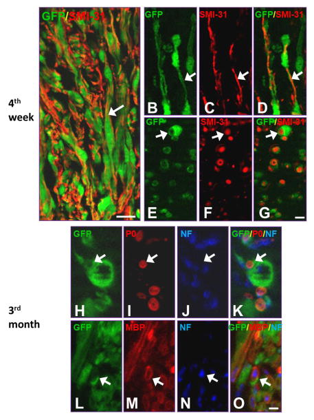
Fig. 6.
Myelination of grafted Schwann cells overexpressing green fluorescent protein (SCs-GFP) post-transplantation. A: Immunofluorescence staining demonstrated that grafted SCs-GFP were in close association with axons growing into the graft at 4 wk post-transplantation (arrow). B–D: A longitudinal section shows that a SC established a one-on-one relationship with a single axon immunostained with SMI-31+ (arrows). E–G: A cross section shows that numerous SCs (arrows) wrapped axons (SMI-31+) within the graft region. H–O: Myelinated axons expressed myelin markers P0 (H–K, arrows) and MBP (L–O, arrows) at 3 mo post-transplantation. Scale bar: A, 40 μm; B–G, 20 μm; H–O, 10 μm.

