
Fig. 2.
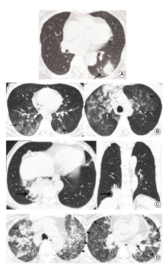
Representative cases for infectious pneumonia. (A) Transverse ultra-low-dose computed tomography (ULDCT) scan in a patient with Aspergillus infection shows a nodule surrounded by a halo of ground-glass opacity in the left lower lobe (arrow). All three observers reached a consensus as an excellent image quality level. (B) Transverse ULDCT scans in a patient with Pneumocystis pneumonia show bilateral patchy areas of ground-glass opacity (arrows). All three observers reached a consensus as an acceptable image quality level. (C) Transverse and coronal ULDCT scans in a patient with streptococcal pneumonia show consolidation involving the posterior basal segment of the right lower lobe (arrows). All three observers reached a consensus as an acceptable image quality level. (D) Transverse ULDCT scans in a patient with coronavirus infection show multiple centrilobular nodules (arrowheads) and bilateral areas of lobular consolidation of peribronchial distributions (arrows). All three observers reached a consensus as an unacceptable image quality level.