
Fig. 3.
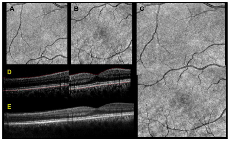
Results of dual spot SSOCT image processing and registration pipeline. Two subfield-of-view volumes were acquired simultaneously, consisting of 1000 A-scans per B-scan for a total of 200 B-scans. The two subvolumes were used to create SVPs, shown for the secondary channel in A and the primary channel in B. The SVPs were then Gabor filtered to accentuate the contrast of the vessels and the peak of the cross correlation was used to obtain the lateral offsets. These offsets were applied and the relative weights of the overlapping region were feathered to generate a registered SVP in C. The inner limiting membrane and the retinal pigment epithelium were automatically segmented, shown in red in D. Using only the region of overlap between the two images, the layer segmentation was used for axial registration, shown after denoising in E.