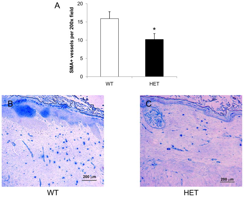
Figure 7.
Analysis of SMA+ vessels in burn wounds on day 21. Immunohistochemical staining for SMA was performed at the healed center of each wound from WT (B) and HET (C) mice and the mean number of stained vessels (± SEM, n = 12 for each genotype) is shown (A). *P<0.05 vs WT, ANOVA with Tukey test.

