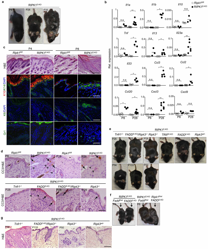
Extended Data Figure 9. Skin inflammation in RIPK1E-KO mice is dependent on RIPK3-mediated necroptosis.
a, Representative macroscopic images of RIPK1E-KO mice. b, qRT–PCR analysis of pro-inflammatory cytokines and chemokines on total skin mRNA from Ripk1fl/fl and RIPK1E-KO mice. c, d, Representative images of skin sections from the indicated mice stained as indicated. In d, arrows point to CC3+ cells and arrowheads depict CC3− dying cells identified by their pyknotic nuclei and eosinophilic cytoplasm. Scale bars, 100 µm in H&E stained; 50 µm in immunostained sections. e–g, Representative macroscopic pictures (e, f) and H&E-stained skin sections (g) of the indicated mice. Scale bars, 100 µm. *P ≤ 0.05, **P ≤ 0.01, ***P ≤ 0.005; NS, not significant.

