
Figure 6. Regional time-course rCBV changes derived from the region-of-interest analysis.
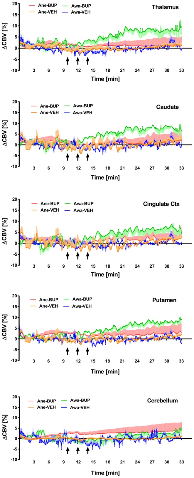
Plots of regional time-course rCBV change (mean ± SEM) derived from region-of-interest (ROI) analyses of the imaging cohort (n = 4) treated with either buprenorphine (BUP at 0.03 mg/kg i.v.) or vehicle (VEH) and imaged under awake (Awa) or anesthetized (Ane) conditions. The black arrows indicate the three individual dosing time periods (30 s drug infusion separated by 1.5-minute intervals), starting at 10-minute after the contrast agent administration. Results indicated that in awake NHP, buprenorphine infusion produces significant increases in rCBV at brain regions with high density of µ-opioid receptors, but not the area with low concentration of µ-opioid receptors (i.e. cerebellum).