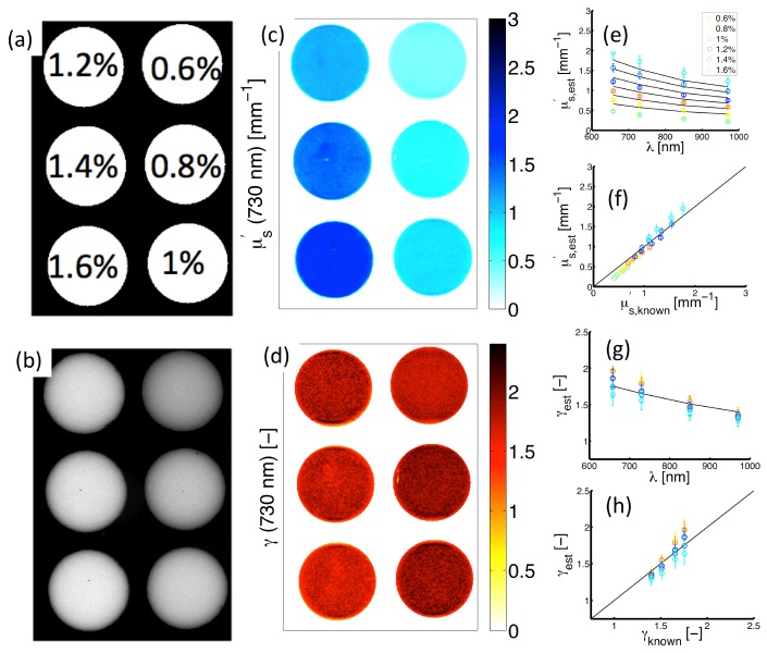
Fig. 5.
Experimental measurements of structured light in Intralipid phantoms. (a) Sampled lipid volume fractions. (b) Diffuse intensity map (). (c) and (d) show spatially-resolved estimates of and at 730 nm. (e) Spectrally resolved in each phantom. (f) Corresponding estimates vs. known values. (g) spectra in each phantom. (h) Corresponding estimates vs. known values.

