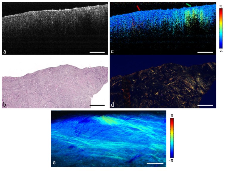
Fig. 4.
Invasive ductal carcinoma replacing the surrounding fibrous environment. (a) Structural OCT image. (b) H&E-stained histology. (c) PS-OCT image (Media 3 (6.3MB, MOV) ). Red arrow indicates tumor, green arrow indicates fibrous stroma. (d) Picrosirius red stained histology. (e) En face PS-OCT projection. Scale bars represent 500 µm.

