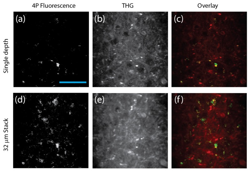
Fig. 3.
4PM and THG images of a mouse brain in vivo. (a-c): 2-frame averaging at a depth of 472 μm below the surface of the brain. (d-f): Average intensity of a 32 μm stack (2 frames per depth, 4 μm step size) ranging from 456 to 484 μm below the surface of the brain. The acquisition time was 4 seconds per frame, and the average power was 23 mW (repetition rate 1.3 MHz) on the brain surface. The scale bar is 50 μm.

