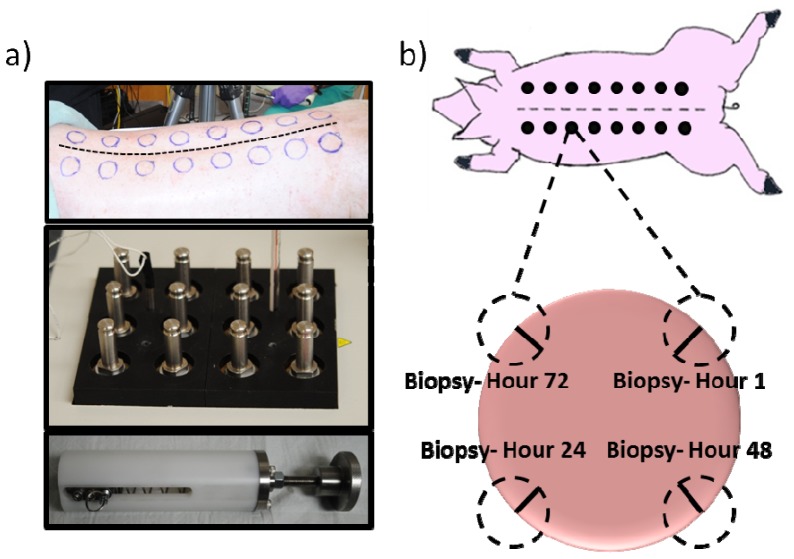
Fig. 2.
Methodology of burn wound creation. a) Top panel shows the dorsum of a pig, with outline for wound spaces delineated. Serrated line shows the spine of the animal. Bottom panels show custom made device made to handle 3cm brass blocks. Spring-loaded device ensures consistent applied pressure. b) Cartoon schematic of the animal highlighting the 3 cm wounds created. For every wound, biopsies (dashed circles) were taken just after burn wound creation (hour 1) as well as hours 24, 48, and 72 post-burn. Solid black lines indicate approximate histological section.

