Fig. 3.
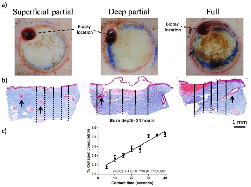
a) Color images of representative burn regions at 24 hours. A prominent red ring representing the zone of hyperemia clearly delineates the edge of the wound bed and the initial 1 hour biopsy punch can be seen in the upper left corner of each wound. b) Trichrome images of burns 24 hours post-injury for 10 second (superficial-partial thickness), 20 second (deep partial thickness) and 40 second (full thickness) burns. Solid black lines indicate collagen coagulation depth, while dashed black lines indicate full thickness of the skin sample. Arrows indicate fully epithelialized hair follicles in superficial burns, while hair follicles in deep partial and full thickness burns are damaged with the epithelium sloughed off. c) Quantification of collagen coagulation depth reveals that contact time correlates well with percent dermal collagen coagulated as determined from histology at 24 hours post injury.

