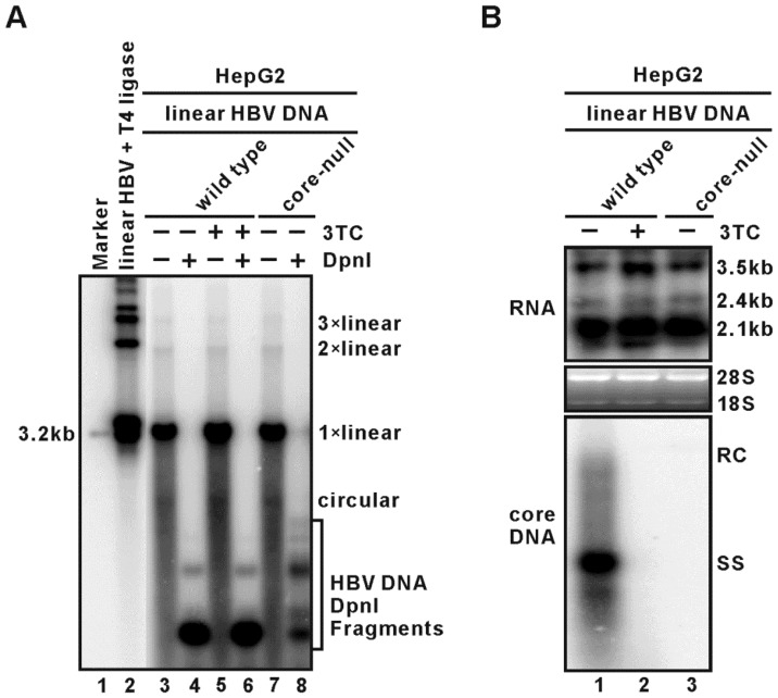
Figure 6. Core protein is not required for HBV transcription in the monomeric linear HBV genome transfection assay.
HepG2 cells were seeded into 6-well-plate and transfected with 4 µg of monomeric linear full-length wild type or core-null HBV DNA for 5 days. One set of wild type HBV linear DNA transfected cells were treated with HBV reverse transcriptase inhibitor Lamivudine (2′,3′-dideoxy-3′-thiacytidine, 3TC) at the concentration of 10 µM. (A) Southern blot analysis of HBV protein-free Hirt DNA recovered from the transfected cells. One set of samples were digested with DpnI before gel loading. Full length linear HBV DNA served as 3.2 kb size marker. In vitro ligation products of linear HBV DNA by T4 DNA ligase served as 3.2 kb DNA ladder. (B) Intracellular HBV RNA and core DNA were analyzed by Northern and Southern blot hybridization, respectively. For RNA analysis, 5 µg of total RNA was loaded on each lane, and ribosomal RNA (28S and 18S) was used as loading controls. The positions of HBV RNA species are marked. For core DNA analysis, half volume of the DNA samples was subjected to electrophoresis. The positions of HBV RC DNA and SS DNA are labeled.

