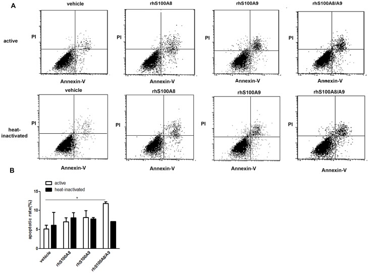
Figure 1. Analysis of PDLC apoptosis using annexin V/PI staining.
PDLCs were treated with 10−6 M rhS100A8, rhS100A9, rhS100A8/A9 or heat-inactivated rhS100A8, rhS100A9, rhS100A8/A9 for 48 h. Apoptosis was then assessed using flow cytometry after annexin V/PI staining. The fluorescence intensity of 10,000 cells was measured and the number of annexin-V-FITC-positive cells was calculated. Statistical analysis revealed that there were significantly more apoptotic cells in the rhS100A8/A9-treated group (10.2475%±2.019%, p<0.05) compared with the control group (5.653%±2.914%). There were no differences between the rhS100A8- and rhS100A9-treated groups. * p<0.05 vs. the control group.

