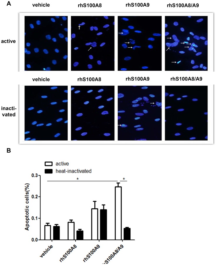
Figure 2. Morphology of PDLCs after DAPI staining.
The morphological changes of PDLCs treated with rhS100A8, rhS100A9, rhS100A8/A9, or heat-inactivated rhS100A8, rhS100A9, or rhS100A8/A9 were evaluated by fluorescence microscopy after DAPI staining. Viable cells displayed a normal-sized nucleus and blue fluorescence, whereas apoptotic cells exhibited condensed chromatin and fragmented nuclei. Treatment with rhS100A8/A9 increased the percentage of apoptotic cells significantly compared with the control (24.5%±5.1% vs. 6.7%±3.1%, p<0.05) and heat-inactivated rhS100A8/A9 (24.5%±5.1% vs. 5.273%±1.14%, p<0.05). Fewer apoptotic cells were observed in rhS100A8-treated cultures whereas more apoptotic cells were present in rhS100A9-treated cells. * p<0.05 vs. the control or heat-inactivated groups.

