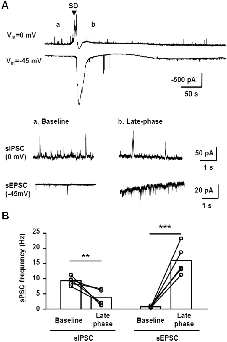
Figure 2. Comparison of sEPSC and sIPSC during SD.
A. Top: representative recordings of pairs of SDs recorded in whole-cell configurations from the same neuron, either at 0 mV or −45 mV. SD onsets were aligned based on DC potential shifts and the onset is indicated by the arrow. Bottom insets show expanded sIPSC and sEPSC recordings during baseline (a) and the SD late phase (b) from the same recordings. B. Quantitative analyses from 5 sets of paired sIPSC and sEPSC recordings, showing frequencies during baseline and the late SD phase. In two cases, recordings from the same neuron could be maintained through two rounds of SD, and in the other three cases a newly-patched CA1 neuron was used for the second recording in the pair. **p<0.01, ***p<0.005, paired t-test, n = 5 each.

