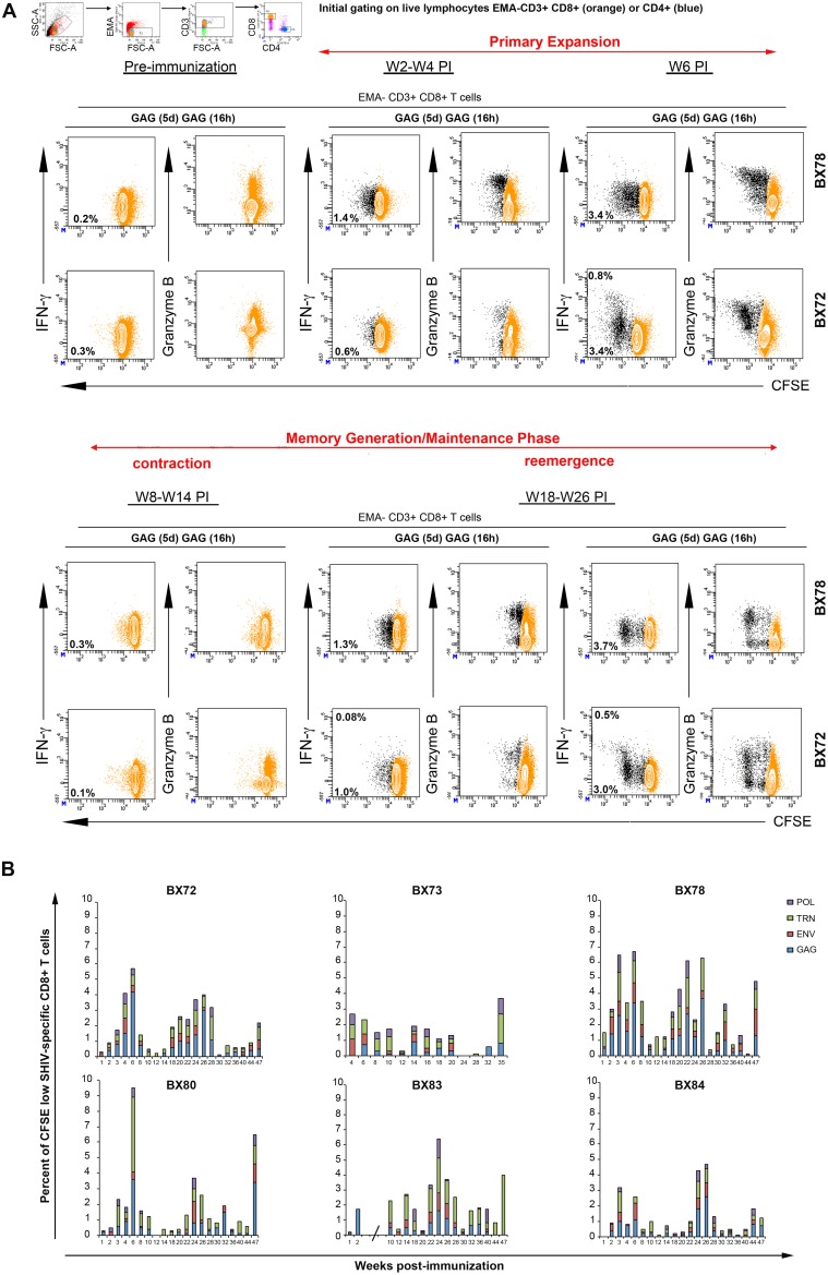
Figure 2. Polyfunctional SHIV-specific CD8+ T cell recall responses.
At various indicated time points pre- and post-immunization PBMCs from vaccinated animals were labeled with CFSE and cultured in presence of specific pools of SHIV peptides Gag, Env, TRN, Pol or medium without peptide for 5 days. On day 5, cells were harvested and restimulated overnight with the same cocktail of peptides (designated as Gag(5d) Gag(16 h)) in the presence of costimulatory Abs and Brefeldin A. Cells were then surface-stained with EMA, CD3, CD8, CD4 mAbs, permeabilized and stained with IFN-γ, IL-2 and Granzyme B mAbs. For flow cytometry analysis, we initially gated on live lymphocytes (low FSC/SSC, EMA-, CD3+), bright CD8+ T cell populations (colored in orange). Antigen-specific T cells were identified by their capacity to proliferate, secrete cytokines and contain lytic molecules (black dots). A) Results for two representative animals (BX72 and BX78) were displayed. The proportion of cells producing IFN-γ (contour plot; upper number) and proliferating (CFSE dilution, contour plot, lower number) in response to specific antigens is indicated in each plot. Frequencies for antigen-specific responses are reported as the percent of cytokine-secreting and proliferating CD8+ T cells after the subtraction of backgrounds obtained with cells cultured for 5 days with medium only and restimulated for 6 h with relevant peptide pools. B) Summary of the frequencies of proliferating (CFSE low) CD8+ T cells detected against each indicated antigen (Gag (blue), Env (red), TRN (green), Pol (purple)) in each immunized animal (BX72, 73, 78, 80, 83, 84) at various weeks post-immunization. Of note, results obtained between W3 and W8 for BX83 were excluded due to non-specific T cell hyperactivation (indicated by an interrupted x-axis).

