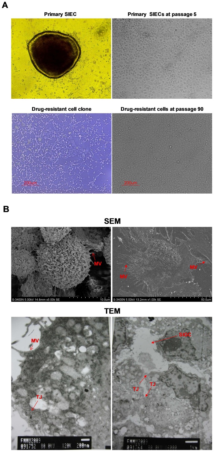
Figure 1. Morphological features of pSIECs and ZYM-SIEC02 cells.
A. Cellular morphology of pSIECs at 7 days in culture (100×) and at passage 5 (100×); a drug-resistant cell clone (100×) and ZYM-SIEC02 cells at passage 90 (100×).No obvious morphological differences were observed between ZYM-SIEC02 cells and primary SIECs. B. Electron micrographs of monolayer cultures of ZYM-SIEC02 cells indicate microvilli (MV) in 3 dimensions andin monolayer culture; tight junctions (TJ) and a small intestine glandular configuration (SIGC) (Red arrow).

