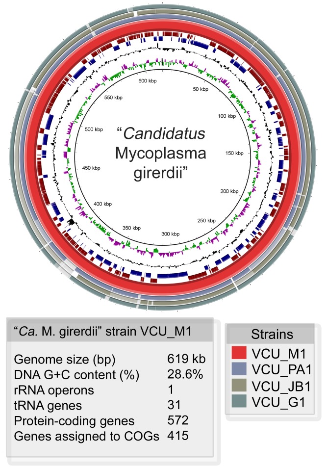
Figure 3. Representation of “Ca. M. girerdii” genomes.
A circular representation of the “Ca. M. girerdii” reference genome (strain VCU_M1) assembled from metagenomic sequences from a mid-vaginal sample. Position 1 is set to the start of the dnaA gene. Outermost circles (1–3) show the alignment (97% or greater identity) of contigs of three different strains from metagenomic assemblies from mid-vaginal samples containing high proportions of “Ca. M. girerdii”. Circle 4 (red) represents the reference strain (VCU_M1). Circles 5 (dark red) and 6 (blue) represent the predicted coding sequences in the forward and reverse orientations respectively. Circle 7 (black) shows the GC content, and circle 8 shows GC skew (pink (-), green (+)).

