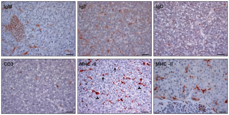
Figure 1. Imunohistochemical detection of different leukocyte populations in trout liver.
Representative photomicrographs of anti-IgM, anti-IgD, anti-IgT, anti-CD3, and anti-MHC-II positive staining in liver sections obtained from unstimulated fish (N = 4). Different phenotypes were observed among MHC-II+ cells including small lymphocyte-like round cells (arrows) and macrophage/dendritic-like cells (arrow heads). Some of these MHC-II+ cells appear as part of the endothelial layer of the blood vessels (asterisks). Counterstained with Mayer's haematoxylin. Scale bar represents 20 µm.

