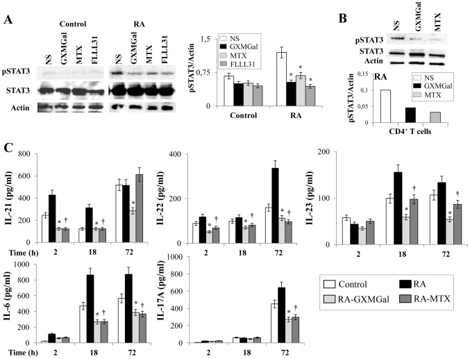
Figure 6. GXMGal effect on Th17 response.
Activated PBMC (A and C) or purified CD4+ T cells (B) (both 5×10⁶/ml) from Control and RA were incubated for 2, 18 and 72 h in the presence or absence (NS) of GXMGal (10 µg/ml), MTX (10 ng/ml) or FLLL31 (5 µM). After 18 h of incubation, cell lysates were analyzed by western blotting. Membranes were incubated with Abs to pSTAT3 and STAT3. Actin was used as an internal loading control. Normalization was shown as mean ± SEM of five independent experiments (A) or as one representative experiment of three with similar results (B). *, p<0.05 (triplicate samples of 5 different Control and RA; RA treated vs untreated cells). Culture supernatants were collected after 2, 18 and 72 h to test IL-21, IL-22, IL-23, IL-6 and IL-17A levels by specific ELISA assays. *, p<0.05 (triplicate samples of 7 different Control and RA; RA GXMGal-treated vs untreated cells); †, p<0.05 (triplicate samples of 7 different Control and RA; RA MTX-treated vs untreated cells) (C).

