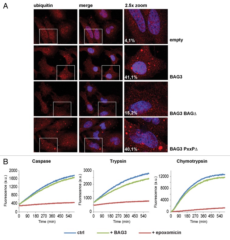
Figure 3. BAG3 induces the accumulation of ubiquitin in cytoplasmic puncta. (A) HeLa cells were transfected with GFP and an empty vector or His-BAG3, His-BAG3-BAGΔ, or His-BAG3-PxxPΔ-encoding vectors. Forty-eight hours post-transfection cells were fixed with 100% methanol for 10 min at −20 °C and subjected to immunofluorescence to investigate the subcellular distribution of ubiquitin. The percentage of cells containing ubiquitin-positive cytoplasmic puncta is indicated. (B) The caspase, trypsin, and chymotrypsin activities of the proteasome were measured in cell lysates from BAG3-overexpressing cells (green line) vs. control cells (blue line) and in cells treated with the proteasome inhibitor epoxomicin (red line) for 30 min. Degradation of the fluorogenic peptide substrates was not impaired in BAG3-overexpressing cells.

An official website of the United States government
Here's how you know
Official websites use .gov
A
.gov website belongs to an official
government organization in the United States.
Secure .gov websites use HTTPS
A lock (
) or https:// means you've safely
connected to the .gov website. Share sensitive
information only on official, secure websites.
