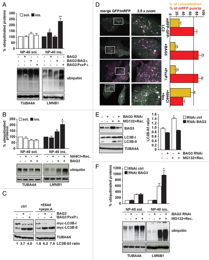
Figure 4. BAG3 is required to induce autophagy and clear aggregate-prone (poly)ubiquitinated proteins following proteasome inhibition. (A) BAG3-PxxPΔ leads to accumulation of insoluble ubiquitinated proteins. HEK293T cells were transfected with either an empty vector or vectors encoding His-BAG3, His-BAG3-BAGΔ, or His-BAG3-PxxPΔ. Forty-eight hours post-transfection NP-40 soluble and insoluble proteins were fractionated. Ubiquitin protein levels were measured in both fractions (*P < 0.05 and **P < 0.001 compared with empty vector; n = 6 independent samples ± sem). (B) HEK293T cells were transfected with either an empty vector or a vector encoding His-tagged BAG3. Twenty hours post-transfection, cells were treated with 20 mM NH4Cl for 6 h and subsequently let recover overnight. The day after, NP-40 soluble and insoluble proteins were fractionated. Ubiquitin protein levels were measured in both fractions (**P < 0.001 compared with empty vector; n = 3 independent samples ± sem). (C) BAG3-PxxPΔ is defective in client rerouting rather than in autophagy induction. HEK293T cells were transfected with either an empty vector or vectors encoding His-tagged FL BAG3 or PxxPΔ. Prior to extraction of total proteins cells were treated for 4 h with 10 µg/ml E64d and 10 µg/ml pepstatin A (+E64d+peps.A) to measure autophagy flux. MAP1LC3B-II/-I ratio (normalized against TUBA4A) was measured. (D) HeLa cells were transfected with mRFP-GFP-MAP1LC3B and either an empty vector, His-tagged FL BAG3 or PxxPΔ. Prior to analysis by confocal microscopy, the cells were treated for 2 h with 20 mM NH4Cl and 0.2 mM leupeptin (+NH4Cl/leu) to block the autophagy flux. Colocalization efficiency of mRFP with GFP signals was measured using ImageJ software and is shown as the percentage of the total number of mRFP puncta that colocalize with GFP puncta (yellow bars). The number of mRFP puncta (that do not colocalize with GFP) in all conditions is also shown as percentage compared with control cells (red bars). The value indicates average and sem from at least 6 images (**P < 0.01). (E) Knocking down BAG3 impairs the compensatory activation of autophagy occurring following proteasome inhibition. HEK293T cells were transfected with either a control siRNA or a specific siRNA for BAG3. Forty-eight hours post-transfection, cells were treated with 10 µM MG132 for 5 h and 30 min and let recover in drug-free medium for 24 h (*P < 0.05 compared with untreated cells; n = 4 independent samples ± sem). (F) BAG3-deficient cells accumulate more insoluble ubiquitinated proteins following proteasome inhibition. HEK293T were treated as described in (C), but recovery was prolonged up to 40 h prior to extraction and fractionation of NP-40 soluble and insoluble proteins. (*P < 0.05 compared with untreated cells; n = 4 independent samples ± sem).

An official website of the United States government
Here's how you know
Official websites use .gov
A
.gov website belongs to an official
government organization in the United States.
Secure .gov websites use HTTPS
A lock (
) or https:// means you've safely
connected to the .gov website. Share sensitive
information only on official, secure websites.
