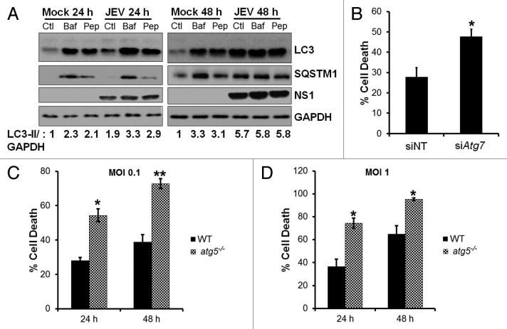
Figure 3. Autophagy-deficient cells are highly susceptible to virus-induced cell death. (A) Mock and JEV-infected (MOI 5, 24 and 48 h), Neuro2a cells were treated with DMSO (Ctl), 100 nM bafilomycin A1 (Baf) for 2 h before fixation or 50 µg/ml pepstatin A (Pep) for 12 h before fixation, and protein extracts were analyzed by western blotting with LC3, SQSTM1, NS1, and GAPDH (loading control) antibodies. (B) Percentage of death in JEV-infected (MOI 1) control/ ATG7-depleted Neuro2a cells at 48 h pi. (C and D) WT (black bar) and atg5−/− (hashed bar) MEFs were infected with JEV at MOI 0.1 (C) and MOI 1 (D), and percentage of cell death was analyzed at 24 and 48 h pi. Presented are mean ± standard error of values obtained from 3 independent experiments. The Student t test was used to calculate P values. *P < 0.05, **P < 0.01.

An official website of the United States government
Here's how you know
Official websites use .gov
A
.gov website belongs to an official
government organization in the United States.
Secure .gov websites use HTTPS
A lock (
) or https:// means you've safely
connected to the .gov website. Share sensitive
information only on official, secure websites.
