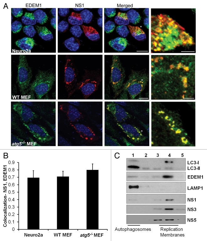
Figure 6. JEV NS1 localizes to EDEM1-positive vesicles. (A) JEV-infected Neuro2a cells, and WT and atg5−/− MEFs (MOI 5, 24 h) were stained with EDEM1 (green) and NS1 (red) antibodies. Color-merged images are shown in the third panel. Panels on the extreme right show magnified view of the region marked with *. Scale bar: 10 µm and 3 µm (extreme right panels). (B) Colocalization of NS1 with EDEM1 in Neuro2a, WT and atg5−/− MEFs (C) Postnuclear supernatants of JEV-infected Neuro2a cells were fractionated on a discontinuous Optiprep gradient. Five fractions were collected from top to bottom of the gradient and probed with antibodies against LC3, EDEM1, LAMP1, NS1, NS3, and NS5.

An official website of the United States government
Here's how you know
Official websites use .gov
A
.gov website belongs to an official
government organization in the United States.
Secure .gov websites use HTTPS
A lock (
) or https:// means you've safely
connected to the .gov website. Share sensitive
information only on official, secure websites.
