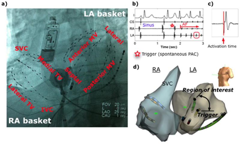
Figure 1.

Catheter placement and recordings of AF initiation. (a) Fluoroscopy showing 64-pole basket catheter in each atrium, implanted ECG monitor (Reveal, Medtronic, MN), catheters in the coronary sinus and LA, and esophageal temperature probe. (b) ECG and intracardiac signals of spontaneous paroxysmal AF after a PAC trigger, with (c) activation time marking of electrogram. (d) NavX shells of both atria indicating the trigger and region of interest, with separation computed from respective (x,y,z) coordinates. (IVC=inferior vena cava, LA=left atrium, MV=mitral valve, RA=right atrium, SVC=superior vena cava, TV=tricuspid valve.)
