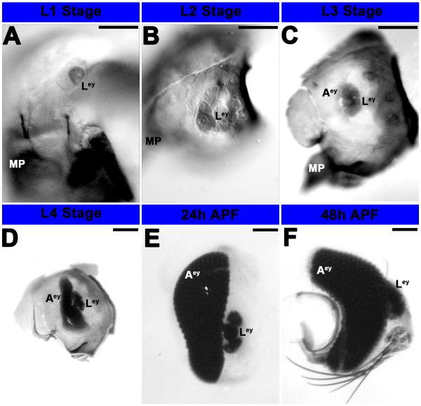
Figure 1. Development of the eye in A. aegypti.
Progressive development of the eye from L1 larvae (A) to 48h APF (F) was visualized through differential interference contrast (DIC) optics (A-F). L1 (A) and L2 (B) larvae possess fairly simple larval eyes (Ley) that are visible at least through 48 h APF; it is unclear if these persist in adults. By stage L3, the adult eye is also visible (C), and it is noticeably larger in L4 animals (D). By the pupal stage (24 h APF in E, 48 h APF in F), additional rows of differentiated photoreceptor cells are visible in the adult eye, which has become a complex primary visual center. Scale bar = 25 microns; Ley: larval eye; Aey: adult eye; MP: mouth parts. Dorsal is oriented upward in all panels.

