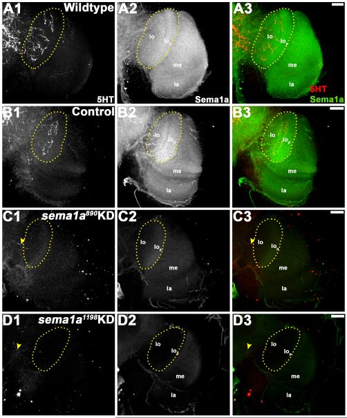
Figure 8. Higher order visual neurons in the optic lobe fail to reach their targets in sema1a KD pupae.
24 h APF wildtype (A1-A3) and control-fed (B1-B3) pupae were labeled with anti-5HT antibody staining, which revealed higher order neural defects in the optic lobe (A1, B1). Similar to larval 5HT neurons, these neurons project from the supraoesophageal ganglion and terminate with a thick arborization in the lobula (lo) region of the optic lobe (highlighted by yellow dots in A1, B1). In comparably staged sema1a KD animals fed with siRNA890 (C1-C3) or siRNA1198 (D1-D3), the 5HT neurons terminate without entering the optic lobe (yellow arrow head in C1, D1). This phenotype correlated with a lack of Sema1a expression in these individuals (Sema1a antibody staining in C2, D2 vs. A2, B2). Overlays of both labels are shown in A3-D3 (5HT in red; Sema1a in green). The optic lobe of one brain hemisegment oriented dorsal upward is shown in all panels. Scale bar = 25 microns; lo: lobula; lop: lobula plate; me: medulla; la: lamina.

