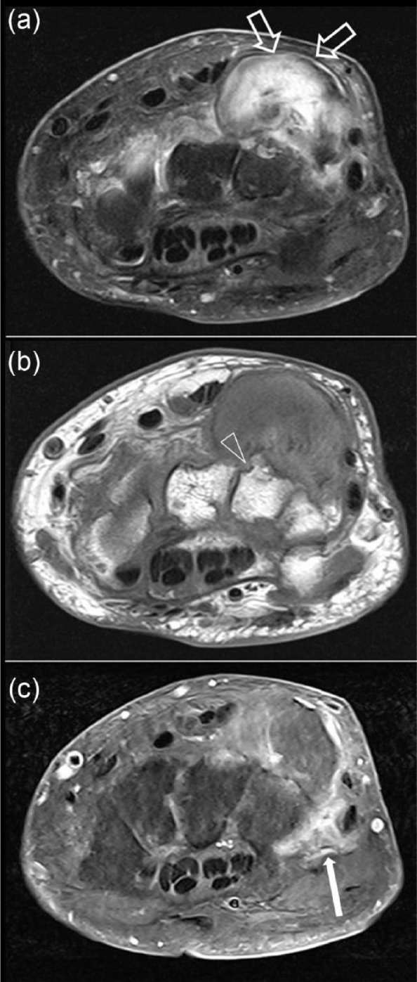
Figure 7.

Magnetic resonance imaging scans of the wrist at the dorsal ulnar aspect: (a) T2 fat-saturated transverse; (b) T1 transverse; and (c) T1 fat-saturated with gadolinium. The scans show a well circumscribed mixed soft tissue calcified mass (open arrow) which represents calcification of tophus with some gadolinium enhancement of soft tissue surrounding the lesion (filled arrow) and periarticular erosions (arrowheads) suggestive of gouty tophus.
