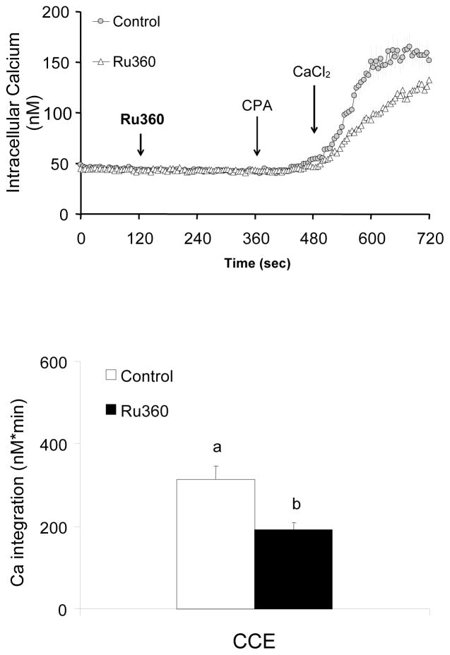
Figure 3. Inhibition of mitochondrial Ca2+ uptake by Ru360 diminished CCE induced by CPA.
Inhibition of mitochondrial Ca2+ uptake by RU360 impaired CCE. Fibroblasts were loaded with Fura 2AM (2 μM) for 60 min. After loading, the media were changed to calcium free BSS, and 3 min later the calcium measurements were initiated. The mitochondrial Ca2+ uptake blocker Ru360 (0, 20 μM) was added 2 min after basal [Ca2+]i measurements, and 4 min later CPA (2 μM) was added. Two min later CaCl2 (final concentration of 2.5 mM) was added. The top panel shows the tracings taken from 50–51 cells. The bottom panel shows the integration of the [Ca2+]i peak over the 3 min interval after calcium addition (495–720 sec). Data are means ± SEM (n=50–51 cells). Different letters indicate values vary significantly (p<0.05) from the other groups by ANOVA followed by Student Newman Keul’s test.

