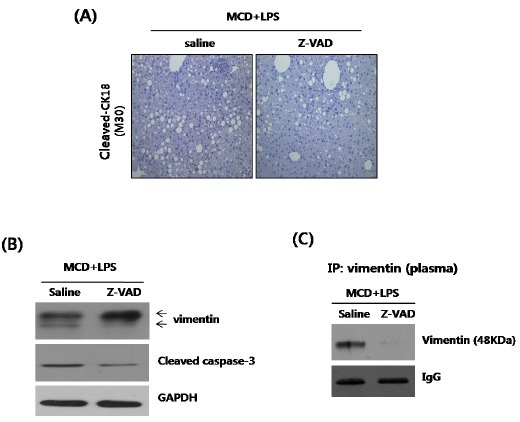
Fig. 4. Effects of zVADfmk in LPS-injected MCD mice. (A) Representative liver tissue sections from LPS-MCD mice injected with either saline or zVADfmk (10 mg/kg) were analyzed for cleaved CK18 (M30), by immunohistochemistry staining (original magnification 200×). (B) Liver tissue extracts were prepared from LPS-MCD mice injected with either saline or zVADfmk. The vimentin and cleaved-caspase-3 were quantified by Western blot, using equal amounts of total liver proteins. Expression levels were normalized, relative to GAPDH. (C) The levels of plasma vimentin were determined by immunoprecipitation. Expression levels were normalized, relative to immunoglobulin G.