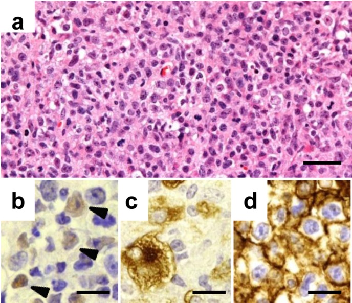
Fig. 2.

Characterization of the LPL in the palpable mass. A mixed population of lymphoid cells is observed in the palpable mass of an affected case (a). The lymphoid cells are positive for EBNA2 (b), LMP-1 (c), and human CD20 (d) by immunohistochemistry. Arrowheads indicate the positive cells in b. Bars=25 µm (a) or 15 µm (b, c, d). Hematoxylin and eosin stain (a) and labeled streptavidin-biotin method (b, c, d).
