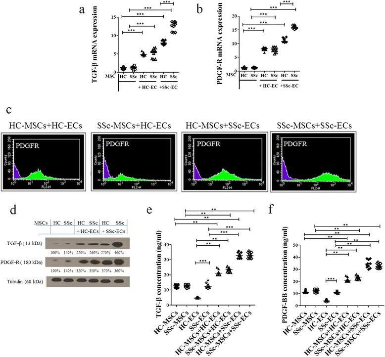
Figure 4.

Mesenchymal stem cells (MSCs) expression of transforming growth factor (TGF)-β and platelet-derived growth factor (PDGF)-R before and after co-culture with epithelial cells (ECs). (a, b) MSCs from patients with systemic sclerosis (SSc) showed greater mRNA expression levels of both TGF-β (a) and PDGF-R (b) compared to the expression levels of MSCs from healthy controls (HCs), when cultured alone. Both SSc- and HC-ECs significantly increased TGF-β and PDGF-R levels in MSCs, compared to the production observed when MSCs were cultured alone, the higher levels observed in SSc-MSC/SSc-EC co-culture (***P <0.0001). Results are expressed as median (range) of triplicate experiments. (c) The cytofluorimetric analysis shows the fluorescence intensity of HC- and SSc-MSCs before co-culture (purple histograms) and after co-culture (green histograms). (d) Western blot analysis of TGF-β and PDGF-R protein in MSCs mirrored the results obtained at molecular level. Pictures are representative of all experiments. (e, f) TGF-β (e) and PDGF-BB (f) ELISA assays. The protein production, in the supernatant mirrored the results observed by qRT-PCR and western blot analyses (**P = 0.0002; ***P <0.0001). Results are expressed as median (range) of triplicate experiments.
