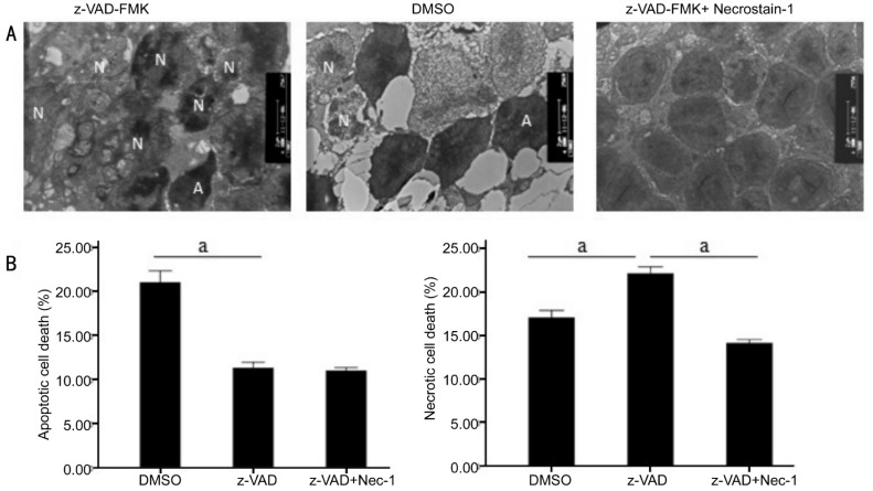
Figure 1. Involvement of autophagosomes formationin necroptosis during RD-induced photoreceptor necroptosis were observed by TEM in three groups (DMSO, z-VAD-FMK and z-VAD-FMK combined with Necrostatin-1 groups).
A: Representative photomicrographs of TEM showed autophagy in the ONL on the third day after RD in the retina (A: Apoptotic cell; N: Necrotic cell). Autophagy formation could be visualized in z-VAD-FMK-treated group, which was by far the most confirmative analysis for autophagy. B: Quantification of necrotic and apoptotic photoreceptor death after RD. z-VAD-FMK treatment increased the percentage of necrotic cells and decreased apoptotic photoreceptor death after RD compared with DMSO treated control groups. z-VAD-FMK combined with Necrostatin-1 substantially led to a decrease in both forms of cell loss. At the same time, autophagy formation was significantly inhibited (n=6, per group, asterisks indicated aP<0.01; Scale bar, 2 µm).

