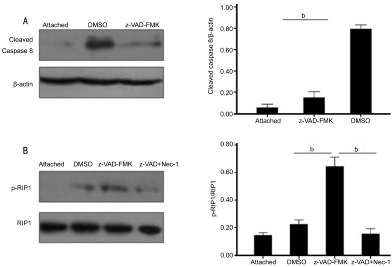
Figure 3. z-VAD-FMK promoted RIP1 phosphorylation by inhibiting caspase-8 activation.
A: Increases in caspase-8 activation after RD and the inhibition of this induction by z-VAD-FMK. Quantification analysis of caspase-8 activation demonstrated a significantly decrease in the z-VAD-FMK-treated retina (0.15±0.02) compared with DMSO-treated retina (0.79±0.02) (n=6, per group, asterisks indicated aP<0.01). B: Immunoprecipitation and Western blotting expression for RIP1 phosphorylation from control group, z-VAD-FMK and DMSO-treated groups three days after RD. Quantification analysis of RIP1 phosphorylation demonstrated a significantly increase in the z-VAD-FMK treated retina (0.64±0.03) compared with DMSO-treated retina (0.22±0.01). Necrostatin-1 combined with z-VAD-FMK treatment substantially inhibited this elevated RIP1 phosphorylation (n=6, per group, asterisks indicated aP<0.01).

