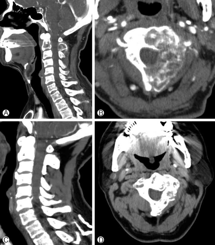
Fig. 2.
(A) Pre-SRS computed tomography (CT) sagittal view, showing an osteolytic lesion infiltrating the C2 vertebral body and spinous process. (B) Pre-SRS axial CT image, showing a tumor mass eroding the left side of the C2 vertebral body, left pedicle, and left lamina. The spinal instability neoplastic score was 10 points. Whole vertebral body involvement rate was 67% and vertebral body osteolysis rate was 65%. (C) Post-SRS CT sagittal view at 24 months follow-up. Cervical spine alignment was good and bone healing was seen at the site of osteolyitc lesion. (D) Post-SRS CT axial view at 24 months follow-up. Bone remodeling was apparent.

