
Figure 2.
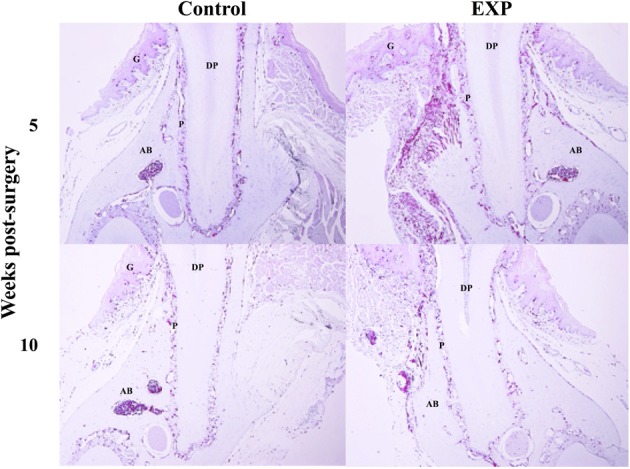
Time-course observations of BMCs around periodontal tissue defects. Experimental periodontal tissue defects were introduced at the buccal surface of mandibular first molar roots in bone marrow chimeric mice. Five weeks after surgery, the number of GFP-positive cells was significantly elevated around the defects. Ten weeks after surgery, GFP-positive cells around the periodontal tissue defects decreased to control levels. G, gingiva; AB, alveolar bone; P, periodontal ligament; DP, dental pulp; D, dentin.