
Figure 3.
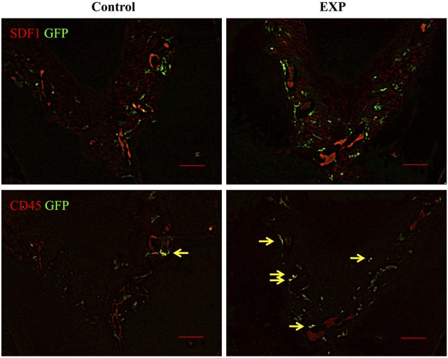
Double staining of GFP and SDF-1/CD45 in periodontal ligaments. In control sites, blood vessel-like structures showed intense staining of SDF-1. GFP-positive cells were observed around SDF-1-positive cells. In experimental sites, SDF-1 expression was ubiquitously detected in periodontal ligament 5 weeks after surgery. The number of GFP-positive cells in experimental tissues was higher than in controls. In experimental tissue, the number of GFP(+)/CD45(+) cells (yellow arrows) was elevated, accounting for 13% of GFP-positive cells (n = 4).