
Figure 1. Immunohistochemical Staining of Primary Striatal Cultures for GFAP and NSE.
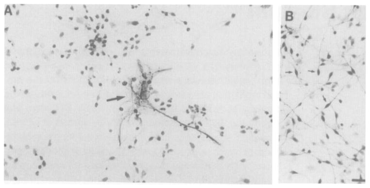
Arrow in (A) points to typical astrocyte stained with anti-GFAP; the surrounding smaller cells are counterstained. Small arrows in (B) point to typical neurons stained with anti-NSE; no counterstain was used. Bar, 20 μm.