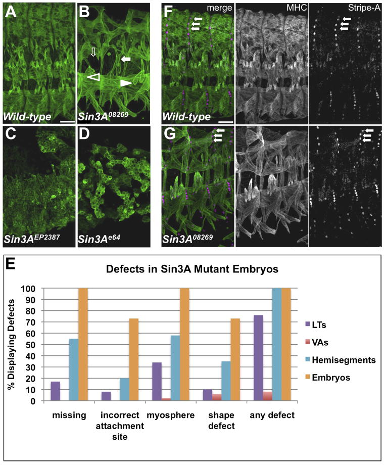
Figure 3. Mutant Phenotypes of and Identity Gene Expression in Sin3A Mutant Embryos.
(A–D) Wild-type (OreR) and Sin3A homozygous mutant embryos stained for MHC. Scale bar, 25 μm. In panel (B), mutant phenotypes are indicated by filled arrows (misshapen), open arrows (missing muscles), filled arrowheads (misattached muscles) and open arrowheads (unattached myospheres). (E) Quantification of mutant phenotypes in Sin3A08269 homozygous embryos. Five abdominal hemisegments from at least 10 embryos for each genotype were quantified. (F–G) Wild-type and Sin3A08269 embryos stained for MHC (white in single channel, green in merge) and StripeA (white in single channel, magenta in merge). Arrows point to SrA-expressing tendon cells. See also Figure S5.

