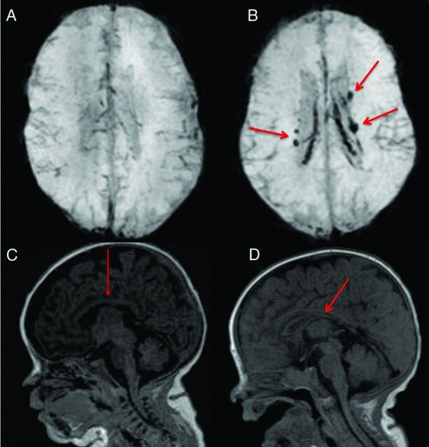
Fig 1.
Examples of conventional MR imaging findings: A, SWI shows brain without blood deposition in an ELBW infant; B, SWI shows brain with blood deposition (arrows show the old blood product) in an ELBW infant with sonography IVH diagnosis; C, normal thickness of corpus callosum (arrow) in an ELBW infant; D, moderate thinning of corpus callosum (arrow) in an ELBW infant.

