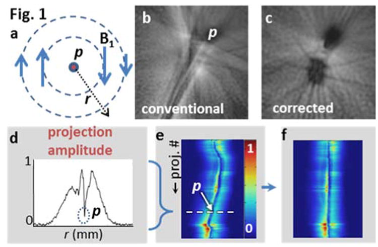
Figure 1.

(a) Transverse field of a loopless antenna detector p shows decreasing B1 with r and azimuthal variation in phase. (b) MRI of an orange shaken ± 3mm (2D radial GRE; 200 spokes spanning 180°; 250μm in-plane resolution; TR/TE=15/6 ms) shows debilitating motion artifacts. (c) Projection shifting all but removes streaking, revealing the fruit's underlying structure. A 1/r intensity filter has been applied to aid visualization. (d-f) The motion correction algorithm consists of re-aligning every azimuthal projection on p.
